High-Quality ASD Closure (Repair) Surgery Cost in India
- ASD Closure Surgery cost in India ranges between USD 4900 to USD 5500.
- The stay in the hospital is for 10 days and 10 days outside the hospital.
- Depending on complexity of disease, the success rate of ASD Closure Surgery is 98.5%.
- Tests required before ASD Closure Surgery are Chest X-Ray, Physical Examination, Electrocardiogram (ECG ) and Echocardiogram.

Brief Overview
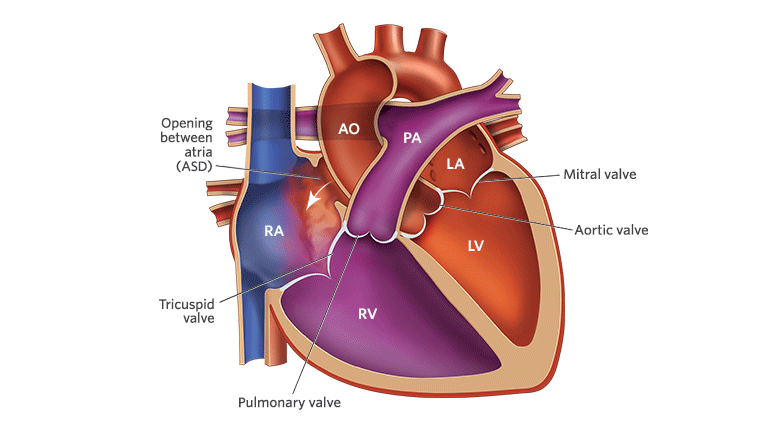
An atrial septal defect (ASD) is a hole in the wall (septum) between the two upper chambers of your heart (atria). The condition is present at birth (congenital).
Small defects might be found by chance and never cause a problem. Some small atrial septal defects close during infancy or early childhood.
The hole increases the amount of blood that flows through the lungs. A large, long-standing atrial septal defect can damage your heart and lungs. Surgery or device closure might be necessary to repair atrial septal defects to prevent complications.
If the ASD is small enough, it can be closed with a special device. The procedure is done in the heart catheterization lab.
What is heart catheterization?
During heart catheterization, the doctor carefully puts a long, thin tube called a catheter into a vein or artery in your child’s neck or groin. The groin is the area at the top of the leg. Then, the catheter is threaded through the vein or artery to your child’s heart.
The doctor who does the procedure is a cardiologist, which means a doctor who works on the heart and blood vessels. This may not be your child’s regular cardiologist.
Signs and Symptoms of ASD
Many babies who are born with atrial septal defects (ASDs) have no signs or symptoms. When signs and symptoms do occur, heart murmur is the most common. A heart murmur is an extra or unusual sound heard during a heartbeat.
Often, a heart murmur is the only sign of an ASD. However, not all murmurs are signs of congenital heart defects. Many healthy children have heart murmurs. Doctors can listen to heart murmurs and tell whether they’re harmless or signs of heart problems.
Over time, if a large ASD isn’t repaired, the extra blood flow to the right side of the heart can damage the heart and lungs and cause heart failure. This doesn’t occur until adulthood. Signs and symptoms of heart failure include:
- Fatigue (tiredness)
- Tiring easily during physical activity
- Shortness of breath
- A buildup of blood and fluid in the lungs
- A buildup of fluid in the feet, ankles, and legs
How the heart works with an atrial septal defect
A large atrial septal defect can cause extra blood to overfill the lungs and overwork the right side of the heart. If not treated, the right side of the heart eventually enlarges and weakens. The blood pressure in your lungs can also increase, leading to pulmonary hypertension.
There are several types of atrial septal defects, including:
- Secundum. This is the most common type of ASD and occurs in the middle of the wall between the atria (atrial septum).
- Primum. This defect occurs in the lower part of the atrial septum and might occur with other congenital heart problems.
- Sinus venosus. This rare defect usually occurs in the upper part of the atrial septum and is often associated with other congenital heart problems.
- Coronary sinus. In this rare defect, part of the wall between the coronary sinus — which is part of the vein system of the heart — and the left atrium is missing.
Risk factors
It’s not known why atrial septal defects occur, but some congenital heart defects appear to run in families and sometimes occur with other genetic problems, such as Down syndrome. If you have a heart defect, or you have a child with a heart defect, a genetic counselor can estimate the odds that future children will have one.
Some conditions that you have during pregnancy can increase your risk of having a baby with a heart defect, including:
- Rubella infection. Becoming infected with rubella (German measles) during the first few months of your pregnancy can increase the risk of fetal heart defects.
- Drug, tobacco or alcohol use, or exposure to certain substances. Use of certain medications, tobacco, alcohol or drugs, such as cocaine, during pregnancy can harm the developing fetus.
- Diabetes or lupus. Having diabetes or lupus might increase your risk of having a baby with a heart defect.
Atrial Septal Defect Complications
A small atrial septal defect might never cause any problems. Small atrial septal defects often close during infancy.
Larger defects can cause serious problems, including:
- Right-sided heart failure
- Heart rhythm abnormalities (arrhythmias)
- Increased risk of a stroke
- Shortened life span
Less common serious complications may include:
- Pulmonary hypertension. If a large atrial septal defect goes untreated, increased blood flow to your lungs increases the blood pressure in the lung arteries (pulmonary hypertension).
- Eisenmenger syndrome. Pulmonary hypertension can cause permanent lung damage. This complication, called Eisenmenger syndrome, usually develops over many years and occurs uncommonly in people with large atrial septal defects.
Diagnosis and Tests for ASD
Hearing a heart murmur during a checkup might cause your or your child’s doctor to suspect an atrial septal defect or other heart defect. For a suspected heart defect, your doctor might request one or more of the following tests:
- Echocardiogram. This is the most commonly used test to diagnose an atrial septal defect. Sound waves are used to produce a video image of the heart. It allows your doctor to see your heart’s chambers and measure their pumping strength.
This test also checks heart valves and looks for signs of heart defects. Doctors can also use this test to evaluate your condition and determine your treatment plan.
- Chest X-ray. This helps your doctor see the condition of your heart and lungs. An X-ray can identify conditions other than a heart defect that might explain your signs or symptoms.
- Electrocardiogram (ECG). This test records the electrical activity of your heart and helps identify heart rhythm problems.
- Cardiac catheterization. A thin, flexible tube (catheter) is inserted into a blood vessel at your groin or arm and guided to your heart. Through catheterization, doctors can diagnose congenital heart defects, test how well your heart is pumping, check heart valve function and measure the blood pressure in your lungs.
However, this test usually isn’t needed to diagnose an atrial septal defect. Doctors might also use catheterization techniques to repair heart defects.
- MRI. This uses a magnetic field and radio waves to create 3D images of your heart and other organs and bodily tissues. Your doctor might request an MRI if echocardiography can’t definitively diagnose an atrial septal defect or related conditions.
- CT scan. This uses a series of X-rays to create detailed images of your heart. It can be used to diagnose an atrial septal defect and related congenital heart defects if echocardiography hasn’t definitely diagnosed an atrial septal defect.
Minimally Invasive Surgery for ASD repair
- The process performed under general anaesthesia.
- Providers perform the surgery by creating only a little 4-6 cm incision in the chest rather than the massive midline-incision.
- The heart-lung system is utilized allowing the heart to be stopped for the stitching of this patch.
- A gentle retractor is inserted, which softly opens the distance between the ribs, allowing the insertion of technical minimally invasive instruments.
- An endoscope is added which provides a high-resolution picture of the heart along with the ASD.
- Working with this technique patients recover faster, and the minimum scar will be hardly visible after the patient recovers.
After ASD Repair
- The patient hospitalized for 3 to 4 days following the operation.
- The incision place might feel tender numbness, itchiness, tightness around the incision area.
- Following the surgical ASD fix, the key medical concern is the healing of the chest incision.
- The first couple of days in the home should unwind doing quiet activities like sleeping, reading, and watching TV.
- It requires approximately 6 weeks to get a chest incision to heal and ready to go back to regular activities.
Before ASD Closure Surgery
The surgeon provides specific instructions to the patient prior to the ASD closure procedure, discussing risks such as bleeding, infection, or adverse reaction to anesthesia.
Patients also meet with the anesthesiologist prior to the surgery to review their medical history. Patients should not eat after midnight the night before the surgery.
On the day of surgery, the patient arrives at the hospital, registers, and changes into a hospital gown. A nurse reviews the patient’s charts to make sure there are no problems.
The anesthesiologist then starts an IV, and the patient is taken to the operating room, where the surgeon verifies the patient’s name and procedure before any medication is given. Surgery will begin once the patient is under anesthesia.
For pediatric patients: It’s important that children are free from infection – including dental infections – for up to six weeks prior to surgery. Please be sure that your child’s immunization records are made available to your surgeon or the nurse.
During ASD Closure
Before the surgery begins, a cardiologist starts a transesophageal echocardiogram (TEE) so the surgeon can look at the heart structure during surgery.
The surgeon then makes an incision in the breastbone to reach the heart, and the patient is placed on a cardiopulmonary bypass machine – which pumps blood to the body, bypassing the heart and lungs except for the coronary arteries – while the heart is stopped temporarily. An incision is then made in the heart’s right atrium to access the defect.
The patch – either the patient’s own pericardial tissue or a synthetic graft – is then stitched onto the hole in the septum to close it.
The heart is closed with sutures, and the cardiopulmonary bypass machine is removed. Pacing wires are placed temporarily on the heart to prevent heart rhythm abnormalities after the operation. Chest tubes are placed to collect residual blood or fluid in the chest after the surgery, and the skin is closed with stitches or staples.
After ASD Closure
After surgery, patients are taken to the intensive care unit and monitored. Pain is likely, and pain medication is given as appropriate. Patients also are on a respirator and have a breathing tube for the first few hours after surgery.
The length of the hospital stay depends on how quickly a patient recovers and can perform some physical activity.
Frequently Asked Questions About ASD Closure Surgery
Q. How much does ASD Closure Surgery Cost in India?
A. The cost of ASD closure surgery ranges from $3500 – $5500 in India.
Q. What is the percutaneous closure device?
A. A percutaneous closure device is a specialized device used to treat patients with atrial septal defect and patent foramen ovale (PFO). In a catheterization procedure, this device is attached to the end of a catheter which is inserted through a vein in the leg and released at the site of the defect to close the hole.
Q. Is a PFO the same as an ASD?
A. Atrial Septal Defect and Patent Foramen Ovale are both birth defects in the septal tissue present between the left and right atria of the heart. ASD is a heart defect that one is born with due to the failure of formation of septal tissue between the atria while PFO only occurs after birth when the foramen ovale, a flap-like valve, fails to close.
An ASD hole is larger than that of a PFO and therefore, more likely to present symptoms.
Q. How does an atrial septal defect occur?
A. Congenital heart defects occur due to failure or errors in the development of heart during the foetal stage. Certain factors like gene defects, chromosome abnormalities and alcohol and drugs uptake by the mother during pregnancy can increase the risk of ASD in the baby.
Q. Can a hole in the heart cause a stroke?
A. Heart defects like ASD and PFO can possibly lead to a stroke. This can happen because the blood clot or a solid particle in the blood can move from the right side of the heart to the left side through the abnormal opening in the septal wall and then travel to the brain, causing a stroke.
Q. Is ASD life threatening?
A. A small atrial septal defect may never cause any problems because it often closes during infancy. The larger defects can cause a number of complications ranging from mild to life-threatening, including right-sided heart failure and stroke.
Q. Can atrial septal defect be inherited from family?
A. Congenital heart defects appear to run in families and sometimes occur with other genetic problems, such as Down syndrome.
Q. Can a patient live with ASD?
A. If the hole is small, the patient might not have any medical problems or it may even close on its own during childhood. The medium and large sized defects can have significant impact on the performance of the heart and lungs. This is because the oxygen-rich blood from the right atrium leaks into the left atrium with oxygen-poor blood and gets pumped back to the lungs, causing increased work-load for the heart.
The larger opening in the septa can cause serious complications and therefore, it must be closed surgically by an experienced cardiac surgeon. For a smaller ASD defect, cardiac catheterization is typically suggested as the primary treatment. But for a larger ASD defect, heart surgery is considered as the best possible option. Minimally invasive ASD heart surgery in India has a high success rate and is carried out by extensively trained and experienced doctors.
