World-Class Medical
Treatment in India
Save 60–80%
Free expert opinion from Fortis, Apollo & Medanta specialists. Dedicated care manager. Complete travel & visa coordination — at no extra cost.
Get Free Medical Assistance
Free · No obligation · Reply within 2 hours
Treatments We Offer
From complex organ transplants and cardiac surgery to cancer treatment and fertility care — expert surgical teams at India's best hospitals, at 60–90% lower cost than the USA or UK.
Organ Transplant
Life-saving transplants performed by India's most experienced surgical teams.
Cardiac Surgery
Advanced heart procedures with world-class outcomes at a fraction of Western costs.
Orthopaedic Surgery
Robotic and conventional joint replacement surgeries with rapid recovery protocols.
Neurosurgery
Complex brain and neurological surgeries by India's leading neurosurgeons.
Spine Surgery
Minimally invasive and conventional spine procedures for lasting pain relief.
Oncology Surgery
Comprehensive cancer surgery including robotic and HIPEC procedures.
Urology
Robotic and minimally invasive urological procedures with high precision.
Fertility (IVF)
World-class fertility treatments with high success rates and compassionate care.
Vascular Surgery
Endovascular and open vascular procedures by experienced vascular surgeons.
Bariatric & Metabolic Surgery
Safe and effective weight loss surgeries with long-term metabolic benefits.
Network of Top Hospitals
We partner exclusively with NABH and JCI-accredited hospitals — the same international standard as leading US and UK facilities. Every hospital in our network is carefully vetted for surgical outcomes, patient safety, and international patient experience.
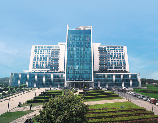
Medanta – The Medicity
Gurugram
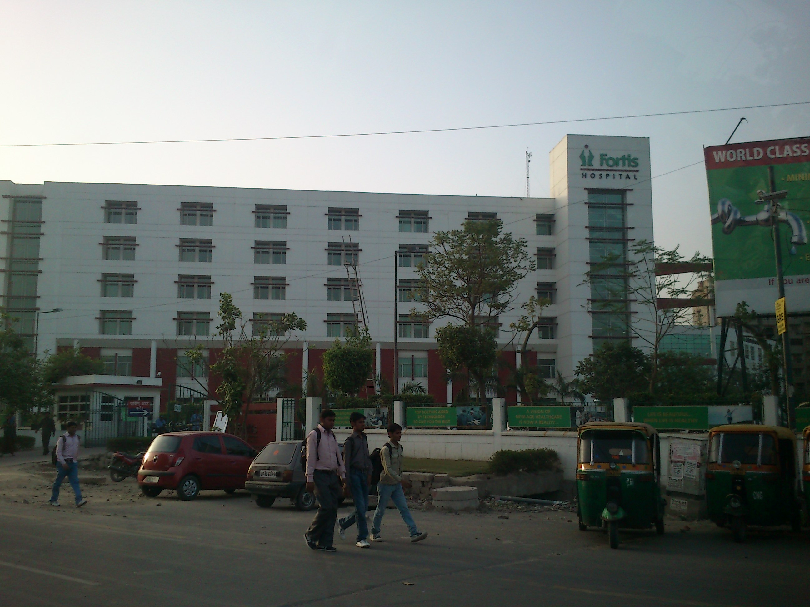
Fortis Memorial Research Institute
Gurugram
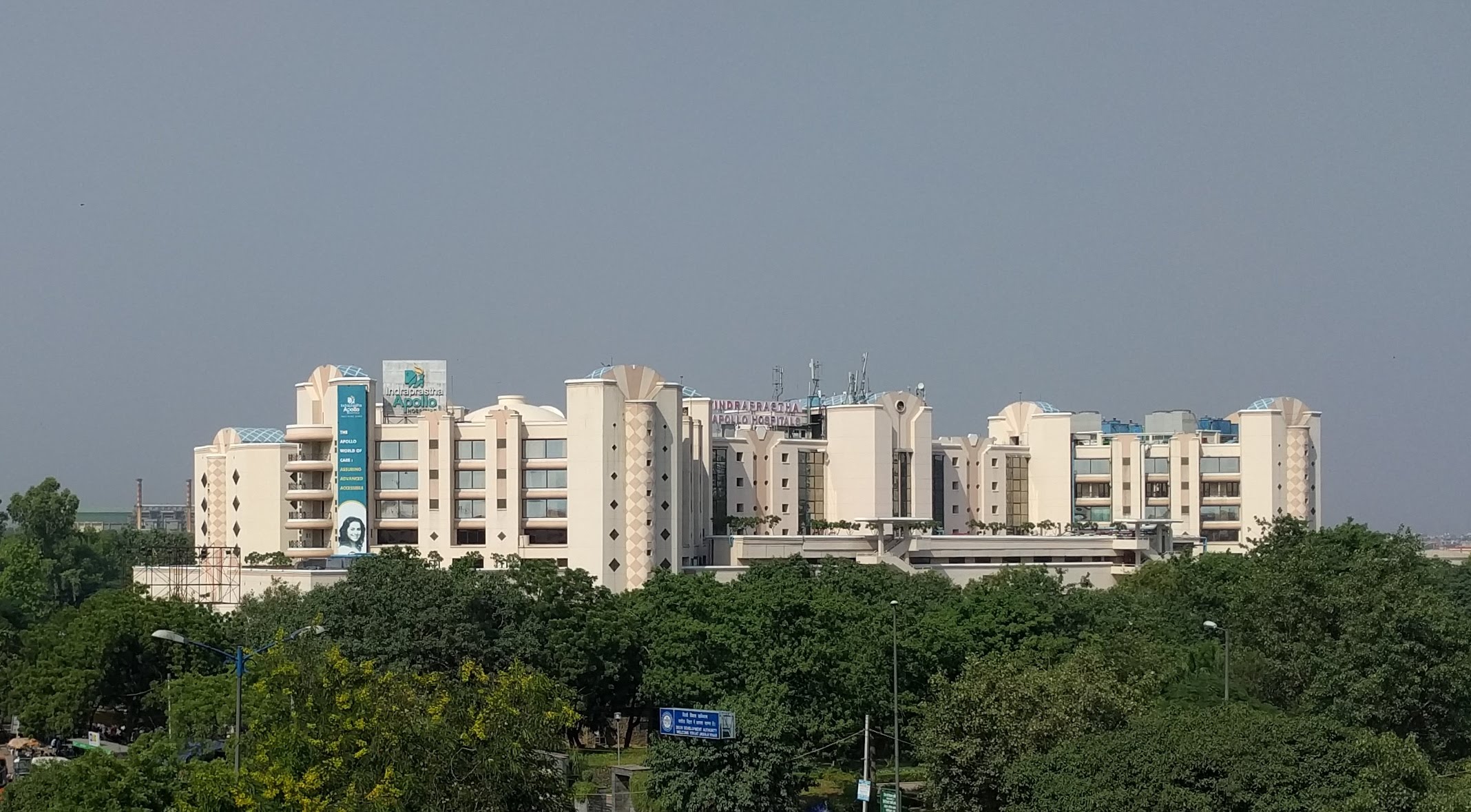
Apollo Hospital
New Delhi
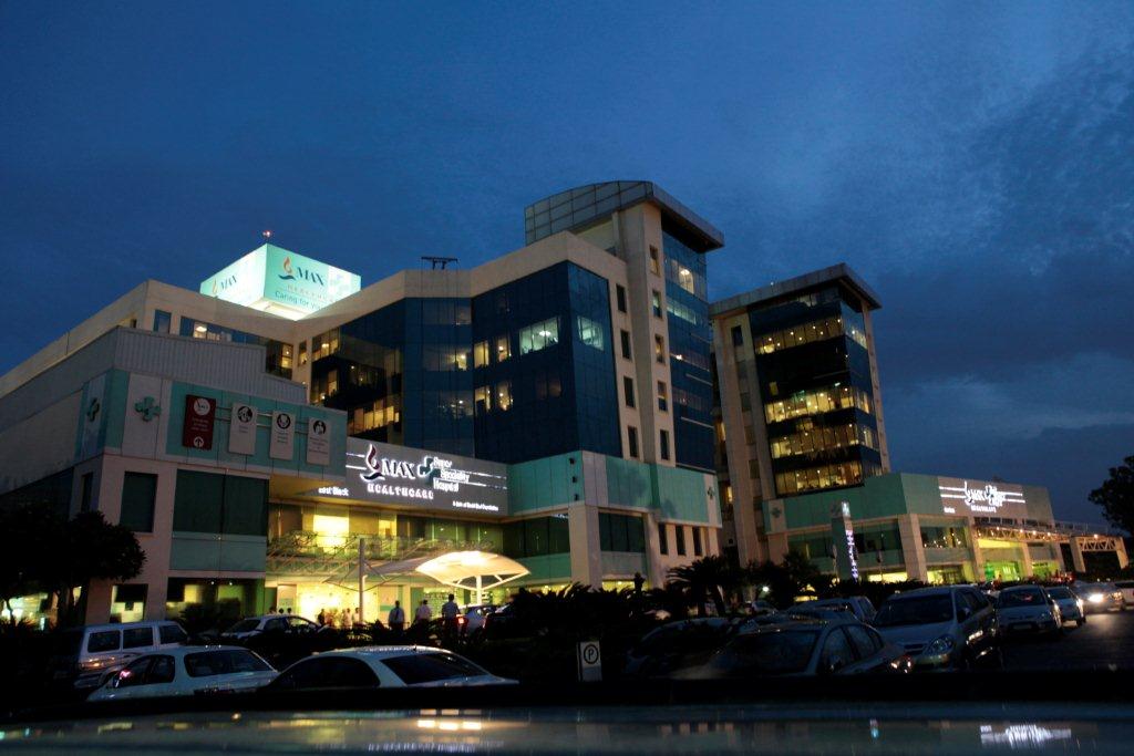
Max Super Speciality Hospital, Saket
New Delhi
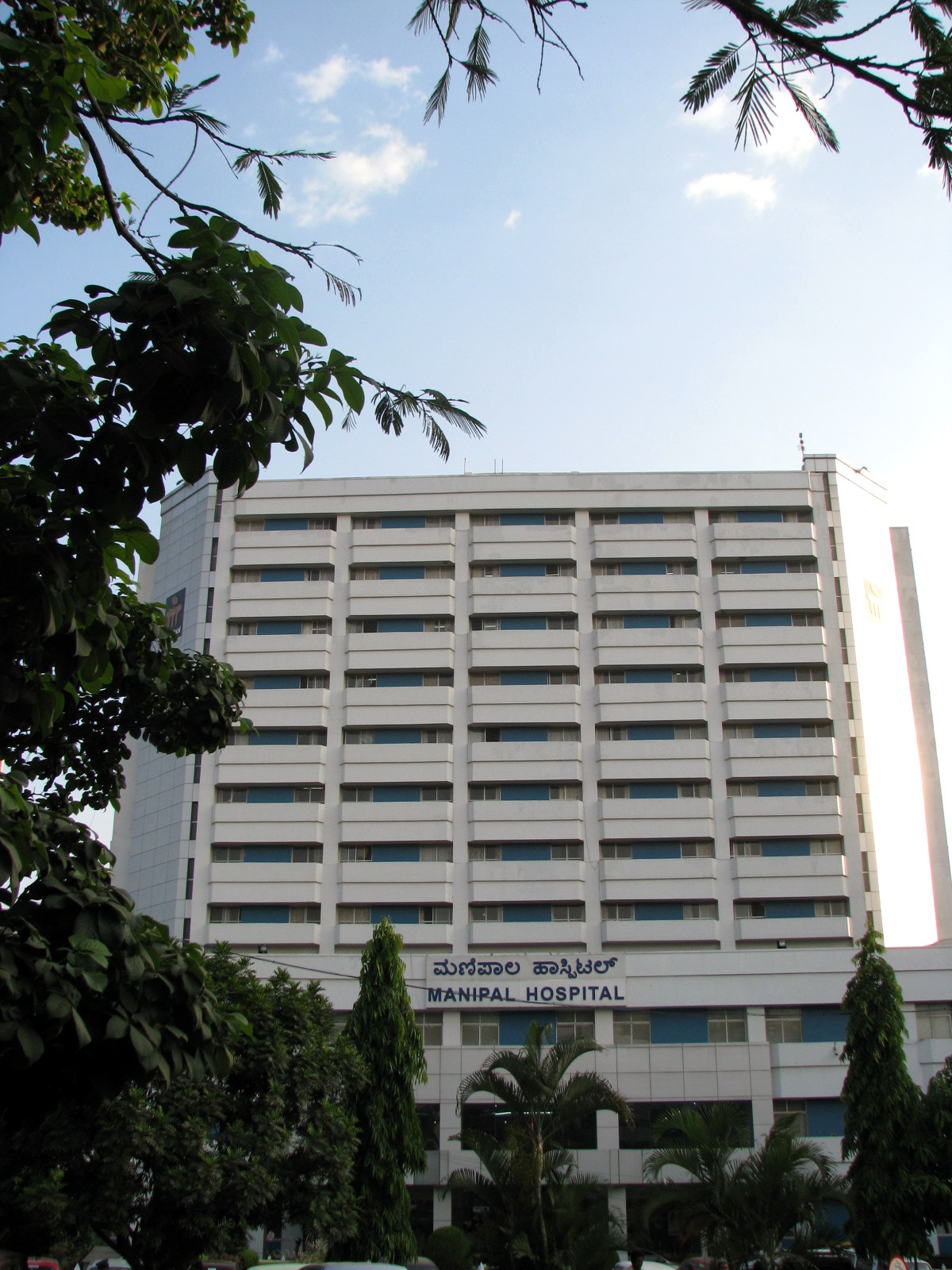
Manipal Hospital (Old Airport Road)
Bengaluru

Kokilaben Dhirubhai Ambani Hospital
Mumbai
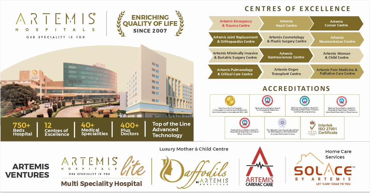
Artemis Hospital
Gurugram
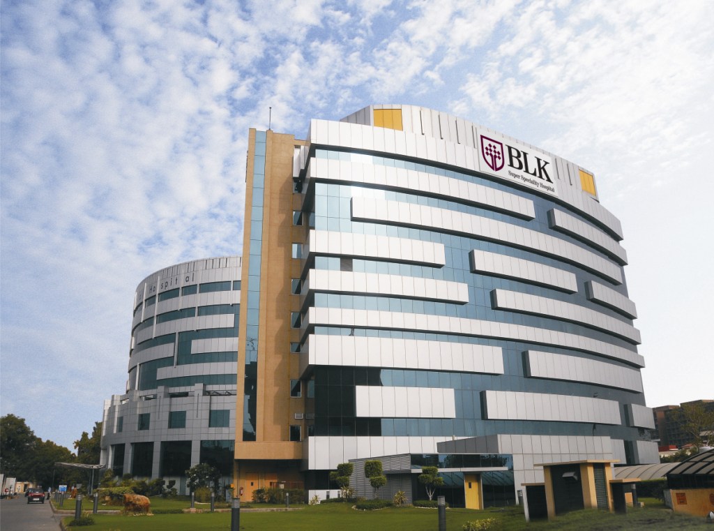
BLK-Max Super Speciality Hospital
New Delhi
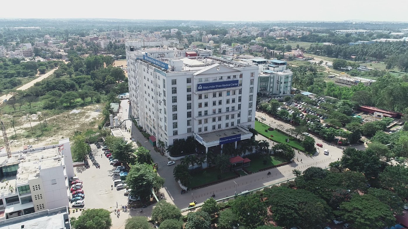
Narayana Health City
Bengaluru
Shalby Hospitals
Ahmedabad
All hospitals in our network carry recognised international accreditations:
Treatment Cost Estimates
India offers world-class surgical care at a fraction of what the same procedures cost in the USA or UK — with no compromise on quality, safety, or accreditation. Below are indicative cost ranges in USD.
Bypass Surgery (CABG)
Cardiac SurgeryTotal Knee Replacement
Orthopaedic SurgeryKidney Transplant
Organ TransplantBrain Tumour Surgery
NeurosurgeryLiver Transplant (LDLT)
Organ TransplantHip Replacement
Orthopaedic SurgerySpinal Fusion Surgery
Spine SurgeryIVF Treatment
Fertility (IVF)Estimates are indicative and based on published hospital rates; final cost depends on individual diagnosis and treatment plan.
All-inclusive packages typically cover surgery, anaesthesia, hospital stay, post-op medication, and dedicated care manager.
International travel, accommodation, and visa fees are not included in the above estimates.
Need a personalised cost estimate for your specific condition?
Request Free Cost Estimate →How Do We Work?
From your first message to your safe return home — we manage every step, so you can focus entirely on your health.
Share Your Medical Reports
Upload or WhatsApp your reports, scans, or diagnosis. Our team reviews everything within 24 hours — at no charge.
Receive Expert Medical Opinion
A specialist at our partner hospital reviews your case and provides a second opinion, treatment plan, and transparent cost estimate in USD.
Visa & Travel Arrangements
We issue a hospital invitation letter for your medical visa, book airport transfers, arrange accommodation near your hospital, and handle your itinerary end to end.
Arrive in India
Your dedicated care manager meets you at the airport, accompanies you to the hospital, assists with check-in, and stays available throughout your entire stay.
Treatment & Recovery
Undergo your procedure at a JCI or NABH-accredited hospital. Your care manager coordinates between you, the medical team, and your family back home.
Return Home & Ongoing Follow-Up
We share your full discharge summary and post-op reports with your home doctor. Remote follow-up consultations are available for 90 days after you return.
Explainer Video
Coming soon — watch how we guide you from enquiry to recovery
Our Services Cover Every Need
We are not a referral agency. We are your full-service medical travel partner — coordinating every step of your journey, at no additional charge beyond your treatment cost.
Free Expert Medical Opinion
Share your reports and receive a specialist's detailed second opinion — including a recommended treatment plan and transparent cost estimate in USD — within 24 hours, at zero cost.
- Response within 24 hours, guaranteed
- Written opinion from a board-certified specialist
- Itemised cost estimate in USD — no hidden fees
Dedicated Personal Care Manager
You are assigned one care manager for your entire journey — from the first video call to your safe return home. They handle every logistics detail so you focus only on getting better.
- Single point of contact — available 24 / 7
- Meets you at the airport; stays throughout your hospital stay
- Coordinates with your family back home in real time
Complete Travel & Stay Coordination
We arrange every logistical detail of your trip — from the official hospital invitation letter for your medical visa to flights, hospital-adjacent accommodation, and all ground transfers.
- Official invitation letter for Indian medical visa
- Flights, hotel & airport-to-hospital transfers
- Translated medical documents & itinerary
Hospital & Doctor Matching
We match your exact condition, budget, and timeline with the right specialist at the right accredited hospital — not a generic referral.
Language Interpretation
Certified interpreters for your hospital consultations in Arabic, French, Russian, Swahili, Bahasa, and 10+ other languages.
Dietary & Cultural Support
Culturally appropriate meals, prayer facilities, local SIM card, and familiar comforts arranged throughout your hospital stay and recovery.
Transparent Cost Estimates
Full, itemised cost breakdowns from our partner hospitals — in USD — before you confirm anything. No surprises, no upsells on arrival.
Insurance Claim Assistance
We prepare and organise all the documentation your international health insurance provider requires for medical reimbursement claims.
90-Day Post-Treatment Follow-Up
After you return home, we coordinate remote video consultations with your surgeon and share all discharge records with your local doctor.
All of the above, from your very first message.
There is no fee for any of these services — MedicareSpots is compensated by our partner hospitals, never by the patient.
Everything You Want to Know
Honest, detailed answers to the questions international patients ask us most. Filter by topic or search below — and if your question isn't here, we're one message away.
We are your end-to-end medical travel partner. From the moment you share your reports, we obtain expert opinions from accredited hospitals, prepare your cost estimate, arrange your medical visa invitation, book your travel and stay, assign you a dedicated care manager on the ground, and stay with you through recovery and follow-up. You make every medical decision — we handle every logistical one.
Yes — completely free. We are compensated directly by our partner hospitals, never by you. Importantly, this does not increase your treatment cost: hospitals offer us pre-negotiated institutional rates that match or beat what you would be quoted booking on your own. Our incentive is your successful outcome, because that is what builds our reputation.
Within 24 hours. Share your reports via WhatsApp (+91 88007 61374) or our enquiry form — pathology results, scans, discharge summaries, anything you have. A coordinator reviews your case, routes it to the right specialist, and comes back to you with next steps and an indicative cost range, all at no charge.
Our partner hospitals — Fortis, Apollo, Medanta, Max, Manipal and others — hold NABH and JCI accreditation, the very same international standards that certify leading hospitals in the USA and UK. Many of our surgeons trained and practised in the US, UK, or EU and perform thousands of procedures every year. We personally vet every facility in our network for outcomes, hygiene, and international-patient care.
Absolutely. We recommend the hospitals and specialists best matched to your specific diagnosis, budget, and timeline — but the final choice is always yours. If you already have a hospital or surgeon in mind, we are happy to coordinate with them directly.
Yes. English is the working language of every partner hospital, and we provide certified interpreters for Arabic, French, Russian, Swahili, Bahasa, and 10+ other languages at no extra cost. Your care manager also stays beside you during consultations to make sure nothing is lost in translation.
That is exactly how we begin. Your free expert opinion includes a specialist’s assessment of your diagnosis and a recommended treatment plan — with zero obligation to proceed. Many patients use it simply to validate advice they received at home before deciding anything.
Most international patients save 60–90% versus US or UK prices, with no compromise on quality. For example: cardiac bypass from $5,000–$10,000 (vs $70,000+ in the US), knee replacement from $5,000–$7,000, and IVF from $2,000–$4,000 per cycle. Every quote we give is in USD and fully itemised.
Your estimate is based on the hospital’s published package rates for your planned procedure and is accurate for a standard treatment course. The only time it changes is if your treating doctor revises the plan after in-person evaluation — for instance, if additional diagnostics or a different procedure is medically required. In that case, you are informed and consulted before any cost is incurred.
Treatment packages typically cover the surgeon’s fees, anaesthesia, operating theatre, hospital stay, standard post-operative medication, and your dedicated care manager. International flights, accommodation outside the hospital, visa fees, and personal expenses are quoted separately so you always see exactly what you are paying for.
You pay the hospital directly — MedicareSpots never handles your treatment funds. Payment is usually made at admission, and hospitals accept international wire transfers, major cards, and foreign currency. We help you understand the schedule in advance so there are no surprises on arrival.
Once your hospital and treatment are confirmed, we issue an official hospital invitation letter for your e-Medical Visa application — typically ready within 1–3 working days. We guide you through the entire online application and tell you precisely which documents you need. The medical visa also permits accompanying attendants.
Yes. India’s medical visa allows up to two attendants to accompany the patient on a linked Medical Attendant Visa. We arrange the invitation letters for them too, and factor their stay into your accommodation and travel planning.
We do — all of it. We book hospital-adjacent accommodation suited to your budget, arrange airport-to-hotel-to-hospital transfers, and help with flights if you wish. Your care manager personally receives you at the airport so you are never navigating an unfamiliar city alone.
It depends on the procedure. A straightforward surgery with recovery may need 2–4 weeks; complex treatments such as transplants can require 6–12 weeks including monitoring. Your specialist’s opinion includes an expected length of stay so you can plan time off and travel for your companions.
Your care doesn’t end at the airport. We provide 90 days of post-treatment follow-up: your care manager coordinates remote video consultations with your treating surgeon, shares your complete discharge summary and reports with your local doctor, and helps you arrange any continuing medication or physiotherapy at home.
Contact your care manager immediately — they remain your direct line to the treating hospital throughout the follow-up period. The surgeon reviews your case remotely, advises your local doctor, and, in the rare event a revisit is needed, we help you re-arrange everything quickly. You are never left to manage a concern alone.
Everything Handled — Enquiry to Recovery
10,000+ patients from 35 countries have trusted us with their most important health decisions. Here's what sets us apart.
- No cost ever
Free Second Opinion
Share your reports and receive expert opinions from JCI/NABH-accredited specialists at Fortis, Apollo, and Medanta — completely free, within 24 hours.
- Beat any price
Lowest Price Guarantee
Direct institutional hospital rates — lower than what you'd pay even if you booked yourself. Full price transparency with no hidden charges.
- Single point of contact
Dedicated Care Manager
One person. On-call 24/7. Coordinates doctors, schedules, reports, and every concern — from your first message through full recovery.
- Stress-free arrival
Full Travel Support
Medical visa invitation letter, airport transfer, hospital-adjacent hotel, local transport, and certified interpreter — all planned and booked for you.
- 3-month aftercare
Post-Treatment Follow-Up
3 months of post-treatment monitoring and coordination. We liaise with your surgeon for follow-up consultations and recovery guidance.
- Always there for you
24/7 Emergency Support
Round-the-clock emergency hotline throughout your trip. Immediate escalation to hospital contacts if any issue arises during treatment or recovery.
Ready to experience the difference?
Get Your Free Quote in 2 Minutes →World-Class Care at a Fraction of Western Costs
All procedures performed by senior specialists at NABH & JCI-accredited hospitals. Prices include surgery, hospital stay, and care manager support.
Don't see your treatment? We cover 200+ procedures. Ask our team →
Get Personalised Treatment Plan →Treatment in the World's Best Medical Hubs
Every destination features internationally accredited hospitals, senior specialists, and full MedicareSpots end-to-end coordination.
India
★ Most PopularJCI & NABH-accredited hospitals — highest volume, lowest prices in Asia.
- Cardiac & Orthopedic Surgery
- Cancer & Oncology Care
- IVF, Transplants & More
Packages from $3,500Get QuoteThailand
Premium ComfortRenowned for 5-star hospital comfort, exceptional nursing & global accreditation.
- Cosmetic & Plastic Surgery
- Dental & Orthodontics
- Fertility & Orthopedic
Packages from $4,500Get QuoteTurkey
Europe's Fastest GrowingStrategic East-West location — world-class surgeons at a fraction of EU costs.
- Hair Transplant (FUE/DHI)
- Bariatric & Weight Loss
- Dental Veneers & Implants
Packages from $4,000Get QuoteUAE
Luxury Medical HubWorld-class hospitals in a luxury destination — full-spectrum specialist care.
- Oncology & Cardiology
- Cosmetic & Aesthetic
- Complex Specialist Care
Packages from $5,000Get QuoteSingapore
Asia's #1 RankedAsia's most advanced healthcare system — unmatched precision and outcomes.
- Cancer & Neurology
- Cardiac & Paediatrics
- Spine & Complex Surgery
Packages from $6,000Get Quote
* Prices are indicative starting figures. Actual cost depends on treatment complexity, hospital tier, and duration of stay. Your dedicated care manager will provide an exact personalised quote.
Your Journey to Treatment, Step by Step
We manage every detail — from your first enquiry to full recovery back home.
Share Your Medical Reports
Send us your reports, scans, or diagnosis via WhatsApp or our enquiry form. Takes under 2 minutes.
Get Free Expert Opinion
Within 24 hours, receive detailed medical opinions and transparent cost estimates from top hospitals.
We Plan Your Full Trip
Your dedicated care manager arranges visa invitation, hospital appointments, hotel, flights, and airport transfers.
Treatment & Recovery
Arrive stress-free. Your care manager stays with you through treatment, hospital stay, and full recovery.
Post-Treatment Follow-Up
3 months of follow-up care after you return home. Remote consultations with your surgeon, anytime you need.
Have a question? Our team responds within minutes. WhatsApp us →
Start My Journey — It's Free →Everything Handled, End to End
From your first enquiry to recovery back home — we coordinate every detail so you focus only on getting better.
Free Medical Second Opinion
Share your reports and get an expert opinion from leading specialists at no cost — before you commit to anything.
Hospital & Doctor Selection
We shortlist the best-matched hospitals and surgeons for your condition from our network of 10 accredited partner hospitals.
Medical Visa Assistance
We issue the official hospital invitation letter for your medical visa and guide you through the full visa application process.
Travel & Accommodation
We book your flights, arrange hospital-adjacent hotels, and coordinate all ground transport including airport transfers.
Dedicated Care Manager
One personal care manager accompanies you through every step — consultations, treatment, hospital stays, and recovery.
Language Interpreter
Certified interpreters provided for your hospital visits, ensuring clear communication with your medical team.
Transparent Cost Estimates
Full cost breakdowns from partner hospitals — in USD — with no hidden fees, before you book anything.
Post-Treatment Follow-Up
3 months of remote follow-up care after you return home, including video consultations with your treating surgeon.
No commitment required · Speak with a medical coordinator
Save 50–80% Versus US & UK Prices
The same world-class surgeons, equipment, and care — at a fraction of Western costs. Here's what that looks like.
| Treatment | US / UK Price | Through MedicareSpots | You Save |
|---|---|---|---|
| Knee Replacement | $50,000 | from $5,500 | 89% |
| Cardiac Bypass | $120,000 | from $10,000 | 92% |
| IVF (per cycle) | $20,000 | from $3,500 | 82% |
| Dental Implant | $4,500 | from $800 | 82% |
| Hair Transplant | $15,000 | from $2,500 | 83% |
* Prices are indicative. Exact quotes provided after reviewing your reports.
Rated 4.9★ on Google. Backed by World-Class Accreditations.
Treatments at India's top hospitals:
Real Patients. Remarkable Results.
10,000+ patients from 35 countries have trusted MedicareSpots. Here are just a few of their stories.
- $165,000 saved
"I was quoted $180,000 in the US. MedicareSpots arranged everything at a JCI-accredited hospital in India for under $15,000. Someone was with me every single day. The outcome was perfect."
- £40,000 saved
"Both knees replaced in one trip. The hospital was more advanced than anything at home. MedicareSpots handled flights, hotel, physio — everything. I couldn't recommend them more highly."
- €13,000 saved
"After two failed IVF cycles in Germany at €8,000 each, we tried India through MedicareSpots. Third time lucky — we're expecting twins! All-in cost was €3,200. The care was exceptional."
Start Your Medical Journey Today —
We Handle Everything
Share your treatment needs and receive a personalised cost estimate, expert medical opinion from top hospitals, and a full travel plan — all completely free.
- Free medical opinion from JCI-accredited surgeons
- Exact cost estimate with multiple hospital options
- Dedicated care manager assigned within 24 hours
- No obligation — decide at your own pace
Get Your Free Quote
Trusted by patients from USA, UK, Canada, UAE & more